
_Mitra Taheri
Taheri is the Hoeganaes associate professor in the Department of Materials Science and Engineering.
The way that electron microscopes work is actually similar to movie projectors: a high-powered beam passes through a material, projecting the image on a screen on the other side. But taking photos of the data can be like trying to project a movie onto a tiny, dirty screen.
A new camera technology developed by Drexel researchers led by Mitra Taheri, Hoeganaes Professor in the College of Engineering, has fixed that. Using a direct detection camera and an image filter, the group can obtain a crisper picture of a material’s chemical structure and composition — and do it much, much faster than before, capturing up to 1,600 frames per second. Plus, the device is sensitive enough so that the microscope can be used to study fragile microbiological samples without damaging them.
Micro_views
Researchers will be able to use the direct detection technology with electron-loss spectroscopy to study biological samples like viruses and bacteria.
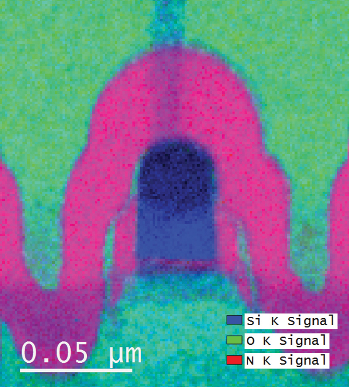
Taheri’s lab uses a Gatan K2 direct detection camera with an electron energy-loss spectroscopy (EELS) microscope — a type that draws its inferences about a sample by measuring how much energy electrons lose when they pass through it. EELS technology is typically used by researchers trying to determine which elements are present in a sample or the chemical structure of one given element.
Drexel is the first to combine the use of these technologies to help researchers collect higher-resolution images of data in a shorter period of time than using a conventional camera — a valuable way to look at the mechanisms behind chemical and physical reactions almost as quickly as they occur.