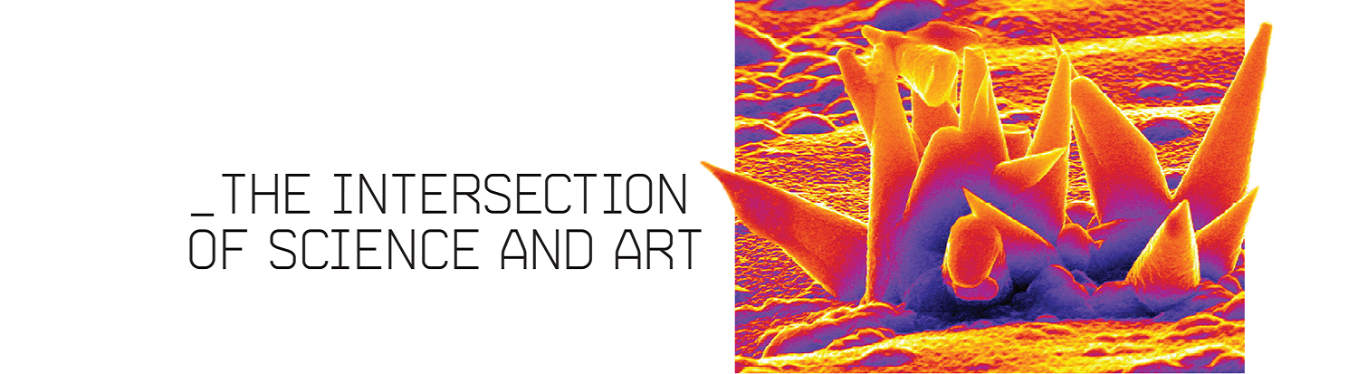
The images that follow were created by researchers and students working in Drexel’s Centralized Research Facilities, using electron microscopes that can achieve magnifications that would scale a grain of sand up to the size of a football stadium. The images, taken at the nano-scale, do not actually have any color; the colorization here was added by Drexel researchers for dramatic effect, creating images that live at the peculiar intersection of science and art.
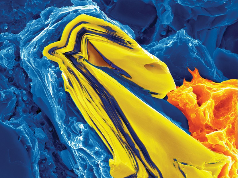
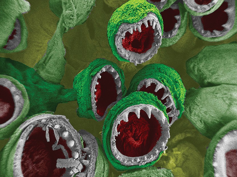
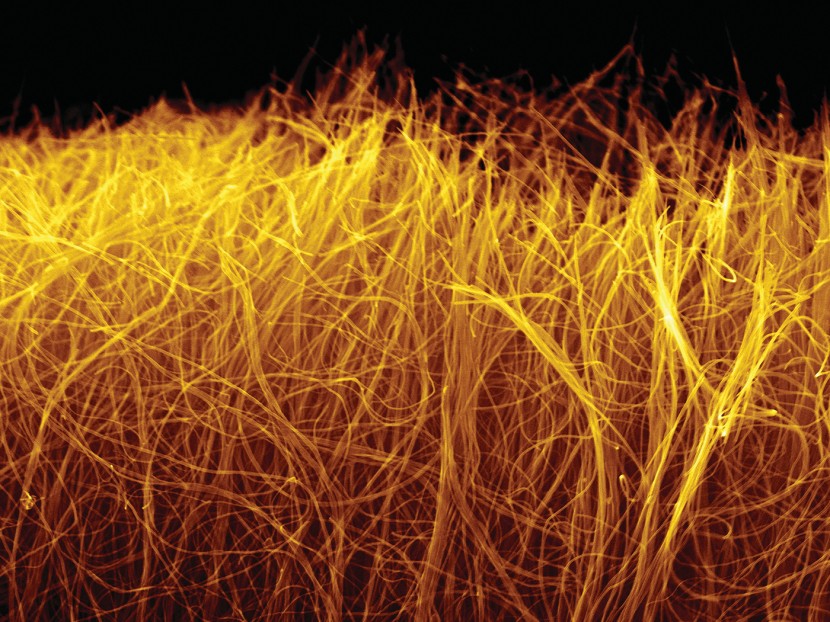
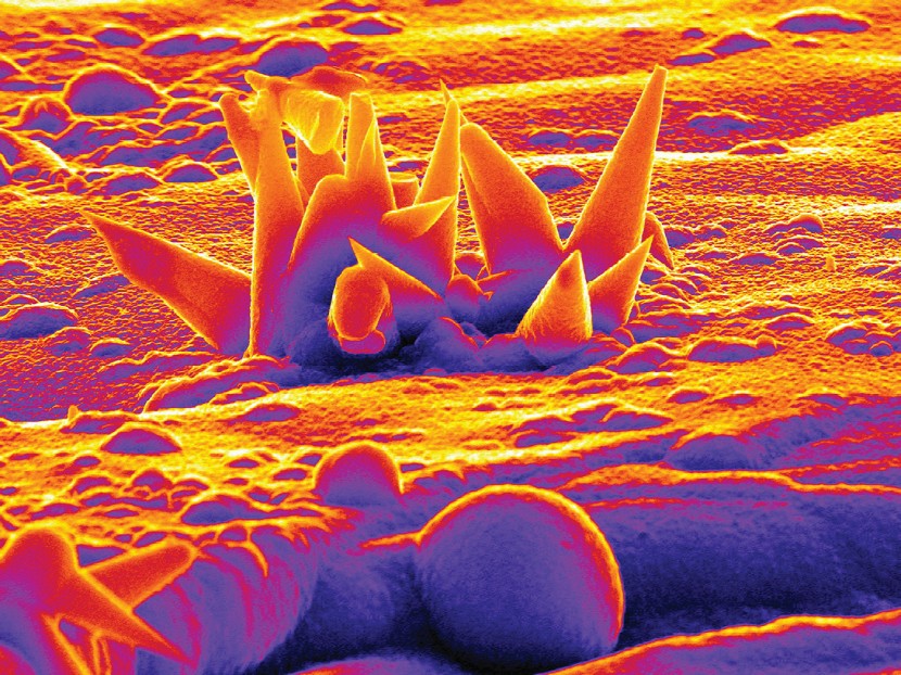

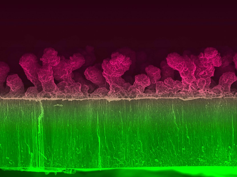
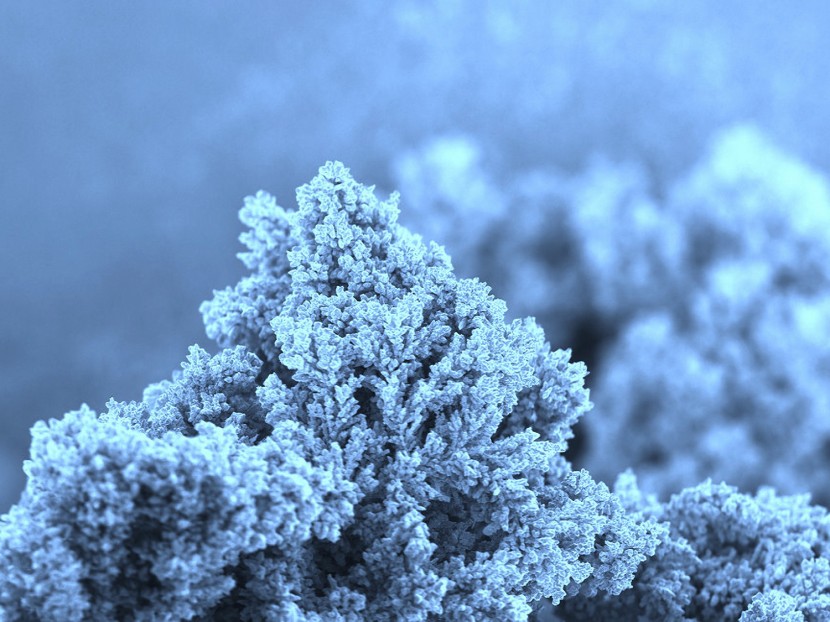
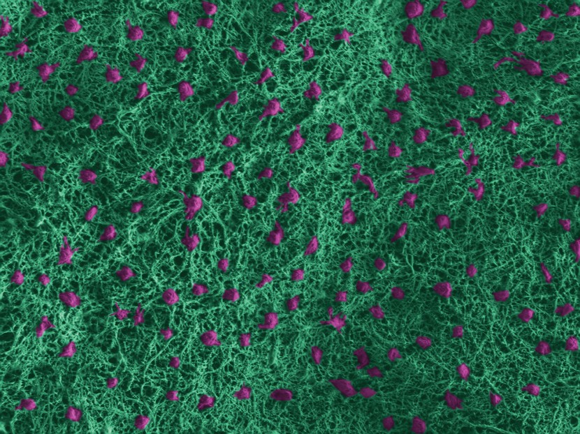

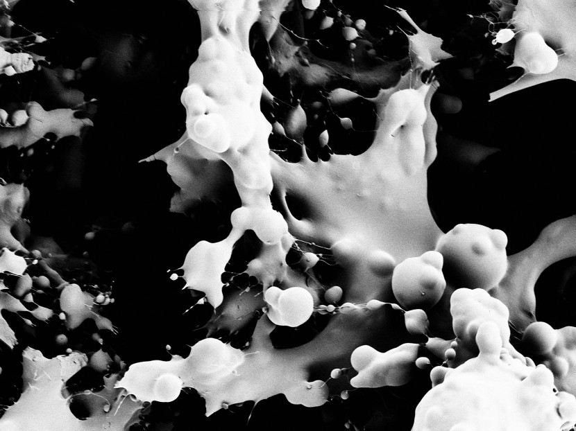
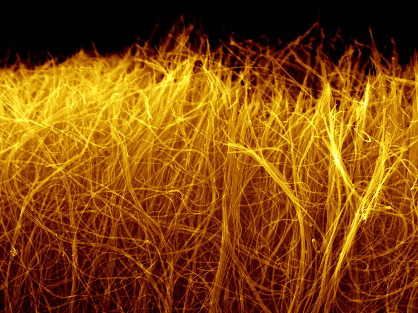
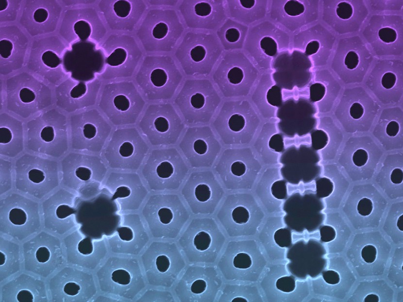
PHOTO 1: Babak_Anasori
MAX Phase Research Group (Michel W. Barsoum)
Fractured surface of a nanocrystalline magnesium matrix composite reinforced with Ti2AlC. A Ti2AlC grain kinked several times during fracture to form a “dragon.” The width of image is 3µm.
PHOTO 2: Jessica_Schiffman
Natural Polymer and Photonics Lab (Caroline Schauer)
Zeiss Supra 50VP SEM, CRF
Fanglike structures that make up a squid tentacle. Each small “mouth” is 400µm in diameter.
PHOTO 3: Min_Heon_and_Boris_Dyatkin
Nanomaterials Group (Yury Gogotsi)
Zeiss Supra 50VP SEM, CRF
Carbon nanotubes in free-standing macroscale sheets (one of a few promising approaches for electric energy storage).
PHOTO 4: Jennifer_S._Atchison
Natural Polymer and Photonics Lab (Caroline Schauer)
Amray 1850 FE, in the MesoMaterials Laboratory
Silicon nanocones grown in a chemical vapor deposition chamber. Tapered semiconductor nanowires have applications in solar cells, antireflective coatings and Li-ion batteries.