

_Craig Johnson
Johnson is director of operations of the Materials Characterization Core of Drexel's Core Facilities.
Inside the glass-faced confines of Drexel’s Bossone Research Enterprise Building on Market Street in West Philadelphia, a top-shelf scientific instrument scans biomaterials and solids, and stretches and tests materials, revealing in spectacular 3D detail the answers to myriad research questions.
It also comes in handy to satisfy a curiosity.
On this day, Lynn Clouser Waddell has come to the building to solve a historical mystery.
Cladding her hands in blue Latex gloves, she opens the lid of a gray archival box. Nestled in white tissue paper sits a stunner — a gold, jewel-encrusted chatelaine timepiece from Drexel’s Founding Collection.
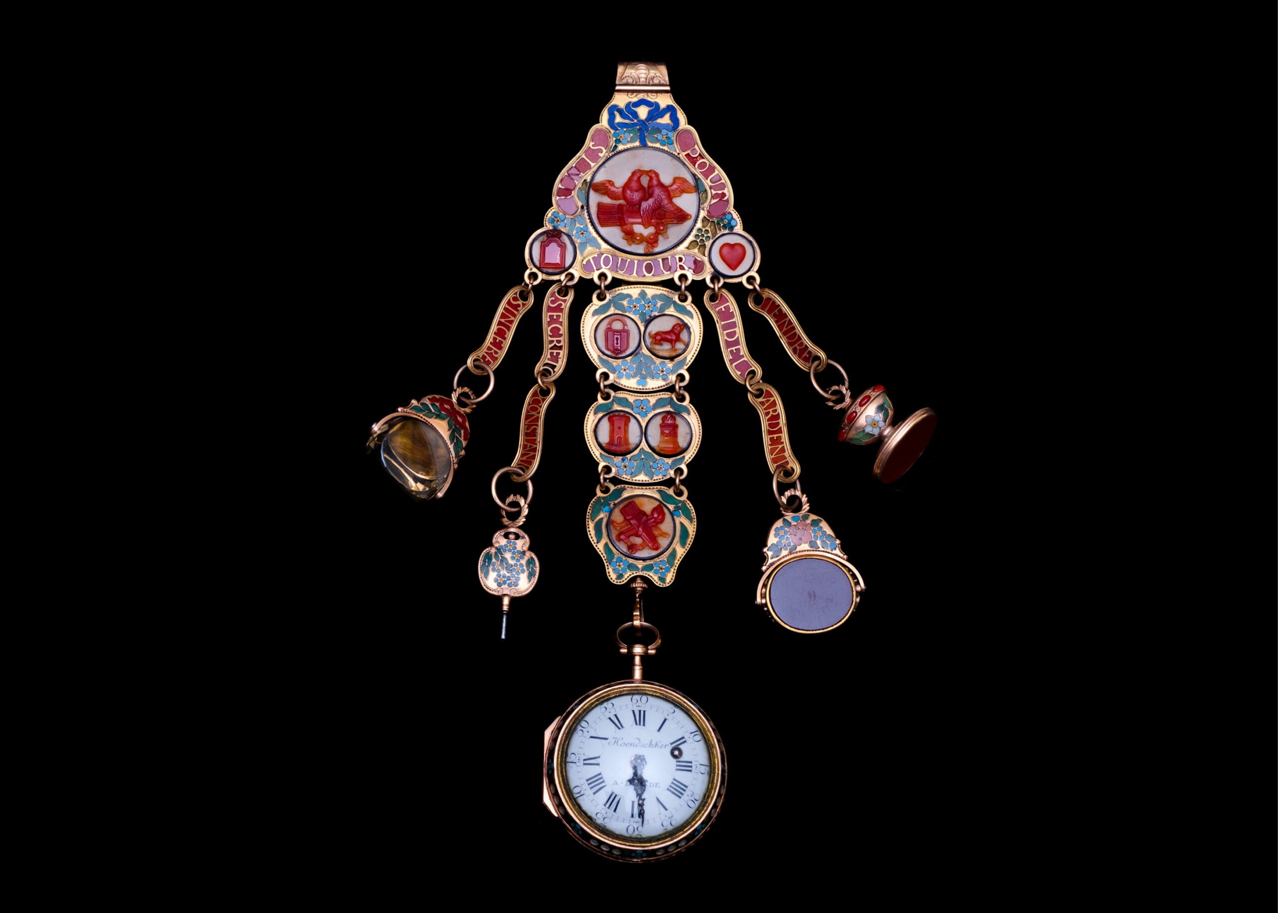
To put Drexel’s 3D nanoCT microscope to the test, researchers scanned a bejeweled timepiece from 1780 believed to have belonged to Marie Antoinette. If there were any markings inside to establish the watch’s provenance, the state-of-the-art microscope would be able to reveal them.
“This one is thought to have been owned by Marie Antoinette,” says Waddell, who oversees the University’s collection of art and antiques. The circa 1780 keychain with a lady’s pocket watch, worth more than $250,000, has bright carnelian cameos, diamond-set hands and an inlay decorated in lapis lazuli, pearls and other semi-precious stones. It was gifted to the University in 1892 by the wife of George W. Childs, a business partner and close friend of University founder Anthony J. Drexel.
Did the chatelaine watch really belong to the queen best known for callous remarks about cake?
Documentation suggests it did. But Waddell would love a no-room-for-doubt answer.
Is there a tell-tale inscription inside the case?
“We haven’t been able to open it,” Waddell says. “It’s jammed. I don’t want to force it.”
That may have been the end of her hunt had it not been for her visit to the suite in Bossone that houses Drexel’s Materials Characterization Core (MCC) lab, one of a number of shared equipment hubs across campus that make up the University’s Research Core Facilities.
The lab recently acquired a $1.65 million X-ray nano-computed tomography (CT) microscope that can provide Waddell with a definitive provenance. The state-of-the-art Zeiss Xradia 620 Versa, which recently replaced a less powerful micro-CT that had been in use since 2006, can peer inside a material or object without damaging it, faithfully revealing tiny internal structures from multiple points of view. Imagine scanning a whole apple and seeing the apple seeds in high-resolution 3D without ever cutting open the fruit.
“The new instrument has many more capabilities,” says Antonios Zavaliangos, the A.W. Grosvenor Professor of Materials Science and Engineering and the principal investigator on the National Science Foundation’s (NSF) Major Research Instrumentation Program grants that helped fund the CT microscope. “With the range of detail we can see, more and more people are interested in using this machine,” he says.
X-ray microscopy is becoming the go-to tool to study an ever-increasing number of materials. Advances in artificial intelligence and machine learning, hallmarks of the next-gen industrial revolution, increase “the need for accurate 3D digitization of material architectures and realistic data-driven computer modeling,” Drexel’s grant proposal points out.
Take the watch. It is the first time that an historical object is being scanned with Drexel’s nanoCT, says Kate Vanderburgh, who was then the research instrumentation specialist responsible for overseeing the instrument and training users.
Drexel’s nanoCT microscope produces thousands of detailed 3D scans. Researchers can rotate solid objects, zoom in on microscopic features, and isolate internal components — without harming or dissembling the object.
“This is our opportunity to see inside,” Waddell adds. “The fact that we have an art collection at an R1 research university does open up possibilities for us to research provenance in a very different way.”
Only a couple dozen universities around the country can boast this specific newer model in their inventory and it’s the only one within 200 miles of Philadelphia, says Craig Johnson, operations director for Drexel’s Research Core Facilities. “An instrument with this kind of specifications is rare,” he says.
Already, the nanoCT, a 6,000-pound, lead-encased chamber the size of an industrial freezer, is finding broad usage. Last year alone, it was reserved for more than 40 hours per week, Johnson says.
Drexel engineers are using the instrument to explore the microstructure of a variety of materials, all with the goal of better understanding why something works or fails. Projects underway are investigating next-generation batteries, novel biomaterials and better pharmaceutical tablets. External clients, such as the University of Pennsylvania and local companies, are scheduling time with it to analyze sea-life specimens and sensors. The Academy of Natural Sciences of Drexel University has used the instrument to help identify new species, understand ocean acidification and shed light on bone fusions through close-up looks at dinosaur bones.
“That’s the amazing thing about this technology,” says Aleister Saunders, Drexel’s executive vice provost for research and innovation. “You can use it in so many different ways. Here we are talking about the benefits to basic research on new materials to our collections, and what does it also tell us about history and craftsmanship?”
“IF YOU JUST TAKE A 2D IMAGE, YEAH, IT’S USEFUL, BUT IT DOESN’T GIVE YOU A COMPLETE PICTURE. THE STRUCTURE IN 3D IS CRITICAL FOR THESE INVESTIGATIONS.”
— LING LI
On this hot August day, Room 106C of the lab is a climate-controlled 72-degrees Fahrenheit. Waddell unhooks the watch from the chatelaine and hands it to Vanderburgh, who will scan it to demonstrate the nanoCT’s power.
“I think it’s fascinating to see beyond what our naked eye can see,” Vanderburgh says, “and how much information it can tell us about a material.”
She wraps a piece of clear packing tape around a block of Styrofoam that is holding the watch upright to ensure it doesn’t wobble during the X-ray.
“Don’t hurt it,” Waddell says, half-jokingly.
“Yeah, no pressure,” Vanderburgh quips. She places the block on the sample holder pedestal and turns toward the machine.
At first glance, it looks rather plain and nothing like the traditional microscope in biology class, this big, white box with a computer monitor to the side and a light on top that turns red to indicate X-ray beams are actively bombarding a sample.
But then, this machine shows what it can do.
Like a medical scan on steroids, the nanoCT has an X-ray so powerful it would burn living tissue, with advanced optical detectors that reveal the target’s internal microstructure down to 350 nanometers. That’s really, really small.
The brother to the nanoCT is the micro-CT, which has a resolution of about five microns, Zavaliangos says. “That is about maybe 14 times smaller than the diameter of a hair,” he says.
The nanoCT significantly ups the game. “We go about 150 times smaller than a diameter of a hair,” he explains.
The new instrument has plenty of other bells and whistles. It can image items as large as a grapefruit. By comparison, the micro-CT that it replaced can only image something up to the size of a fig. Because of the instrument’s wide voltage range (30kV–160kV), it can scan more materials, from polymers and wood to dense metals.
“I haven’t had a sample I can’t see through yet,” Vanderburgh says proudly. That includes iridium, the second-densest material known to humans, she says.
Scientists also can use the nanoCT to study samples in situ. As a device stretches or compresses a material, the machine can capture how the microstructure reacts under stress and over time. Plus, all this work gets done in a fraction of the time the older instrument took, largely because of better detectors.
The scan itself comprises a series of X-ray projections from different angles, each a thin slice of the mounted sample as it rotates and moves up and down between the X-ray source and the detectors. More slices, which can number into the thousands, equate to higher resolution.
Imagine going to a deli counter, Vanderburgh says. “They have a whole hunk of ham, and they slice individual thin slices,” she says. Then the reconstruction software Dragonfly takes the pieces and stacks them together to generate a cool 3D image that can be digitally rotated, pulled apart into segments and zoomed through to explore minute detail. In other words, put together all that lunch meat, and presto! There’s the whole ham.
“It can tell us information about a material at such a fundamental level,” Vanderburgh says, “and give us such a better, global understanding of the entire sample.”
The key is the microstructure, Zavaliangos says. This internal geometry of a material controls its properties. “When we want to produce a new material, or to optimize an existing one, or to understand why a material failed,” he says, “understanding the microstructure is a step of paramount importance. Instruments like the nanoCT and other instruments of the centralized facility are essential tools for the progress that materials science has enabled with discoveries for all parts of our lives.”
Zavaliangos, for one, studies the mechanical properties of solids produced from powders, looking to improve the manufacturing process for pharmaceutical tablets. “We are trying to understand the internal structure of the tablet,” he says. “What happens if you go from one shape to another? What really drives these changes? How can they be minimized or amplified?
“If you do any other technique than tomography,” Zavaliangos adds, “it only shows you what’s going on on the surface.”
Other Drexel researchers are using the technology to probe energy-related devices, biomedical specimens and advanced materials.
A group working with former Drexel chemical engineering professor Vibha Kalra used it to explore novel material architectures to develop more efficient energy storage devices and next-gen batteries. Other faculty are measuring battery behavior during operation and conducting in situ experiments.
Kara Spiller, the URBN Professor of Biomedical Innovation, harnesses immune cells involved in tissue repair to create new biomaterials for regenerative medicine in the growing field of immune engineering. Thanks to the nanoCT’s capabilities, the co-PI on the instrument grant can analyze high-res, 3D images of a mouse model’s whole lungs to understand the effects of a novel treatment for pulmonary fibrosis. “This is important because fibrosis is not uniform,” she says. “We need to analyze the whole lungs.”
The equipment in the MCC is also important to researchers in Drexel’s NanoBiomechanics Lab, which investigates the role of extra-cellular matrix (ECM) biomolecules in the development of diseases such as osteoarthritis. The nanoCT offers critical insight into erosion of cartilage in a mouse model and the promise of engineered hydrogels, information that can dramatically enhance scientific understanding of how cells and tissues interact with implants.
This technique has also demonstrated great potential in the field of nanomaterials. Each week, Bita Soltan Mohammadlou, a mechanical engineering doctoral student in the Drexel Nanomaterials Institute (DNI), schedules time on the nanoCT to investigate the internal structure of MXene-based samples.
Distinguished University and Charles T. and Ruth M. Bach Professor Yury Gogotsi, who directs the institute, co-discovered the versatile 2D nanomaterials with Distinguished Professor of Materials Science and Engineering Michel Barsoum 14 years ago.
In her research, she studies the mechanical properties of MXenes using different characterization techniques. More recently, she has shown the benefits of connecting scanning electron microscopy that analyzes the surface of MXene-coated textiles, composites and aerogels with cutting-edge tomography that sees internal features such as microstructure, distribution and defects. This allows for more precise analysis of materials and ultimately guides optimization and development of their synthesis and applications.
“This is the first time nanoCT has been systematically applied to analyze different MXene systems,” says Soltan Mohammadlou, who has written a paper that highlights the transformative potential of the technology in the study of these 2D nanomaterials. “So, it’s a very advanced and exciting technique to work on. I can’t wait to visualize my data each time.”
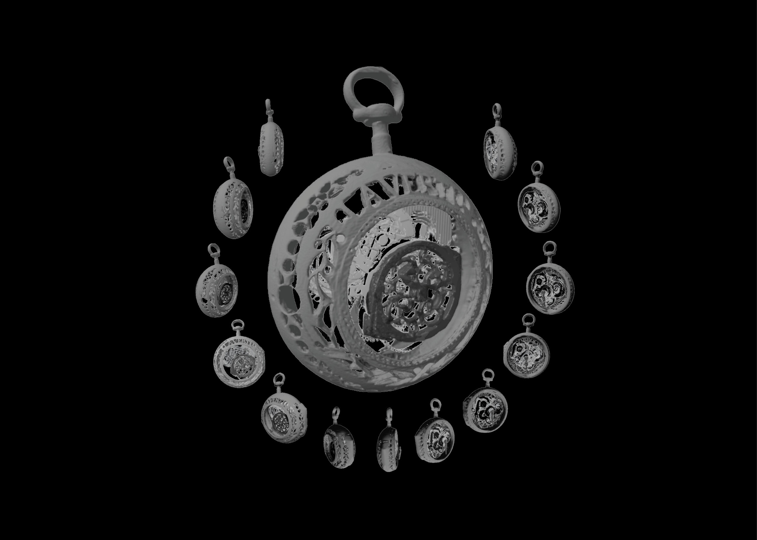
Vanderburgh places the holder with the watch inside the instrument. From the computer, she moves the X-ray source closer to the sample for better resolution and sets the machine to its maximum power of 160 kilowatts, given the object’s high density. Soon, an X-ray of circles layered atop each other appears on the monitor.
“Oh, I see the gears,” says Waddell, her face lighting up. “The mechanism is beautiful.”
No one spots any engraving on the inside. But this is a single image, what Vanderburgh calls a preview to check the positioning of the watch before the complete scan with its many slices takes place. That will show more detail and, fingers crossed, an inscription.
See how Drexel’s advanced materials scanner is able to tour the 360-degree interior of an antique timepiece. Video courtesy of Bita Soltan Mohammadlou.
Certainly, Grace Goetcheus, a doctoral student in the Biodiversity, Earth and Environmental Science (BEES) department and one of the many students trained on the nanoCT, was impressed at the detail when she scanned fossilized tail vertebrae of juvenile dinosaurs. Based at the Academy of Natural Sciences, the 26-year-old from Maryland is searching for clues as to why diplodocids, a group of long-necked sauropod dinosaurs, have a high prevalence of fused vertebrae in their tail — work that could provide insight into human spinal fusions.
Is it, Goetcheus asks, because of normal injuries? Or is it because of the gigantic size of sauropods, the largest terrestrial animals to ever live? “You can see that a fracture occurred externally,” she says, citing the new bone growth around a break known as a callus. “But with the CT machine, you can zoom in to specific, smaller areas and see where the line of the old bone is and where all that new bone has grown to create that external callus.” An examination of the juncture of two fused vertebrae suggested the ligaments ossified, which usually occurs later in life in humans but in this instance was appearing among young sauropods — and potentially as an adaptation to gigantism, she says.
Kyle Luckenbill, collection manager and imaging specialist at the Academy, has been scanning specimens in collaboration with visiting researchers. These scans will be uploaded to Morphosource.org in support of the oVert project, a multi-institution, NSF-funded initiative to digitize vertebrate museum specimens and make the images available at no cost to the public and to researchers. He says that during the process, South American catfish in the collection with widespread geographical ranges that had initially been thought of as the same species were confirmed to have different bone structures, setting them apart from each other. One highlight of the technology, Luckenbill adds, is its non-destructive nature — one needn’t alter the specimen with preparations traditionally used to examine the skeleton.
“It comes out,” he says, “the same way it went in.”
For BEES Associate Professor Jocelyn A. Sessa, the Academy’s associate curator of invertebrate paleontology and a co-PI on the instrument proposal, having an instrument like this in the Mid-Atlantic means saving time and energy. She used to travel all the way to the American Museum of Natural History in New York to access tomography, crucial to her research on the small, delicate sea snail shells that appear to be bioindicators of the ocean’s changing acidity. It hampered her work, she says. Now, she need only go to Drexel’s campus. “This,” Sessa says, “was part of our pitch for the grant.”
As far back as 2015, Zavaliangos was looking to replace the Bruker Skyscan 1172 micro-CT that he and others used often with the latest technology. He submitted proposals twice to no avail. “At some point, I gave up,” he says.
But a colleague, Professor Antonios Kontsos, convinced him to try again, and the third time proved the charm. The $1.23 million, 2022 grant covers 75% of the instrument’s costs with Drexel funding the remainder. “The whole [MCC] facility is full of instruments won through these types of grants,” Zavaliangos says.
Opened in 2006, the 3,500-square-foot MCC houses nine major instruments, including several electron microscopes, X-ray diffractometers, an X-ray photoelectron spectrometer and, of course, the nanoCT. Each has a sweet spot. The X-ray microscope handles a centimeter to just under a micron, while the scanning electron microscope covers 1 mm to a few nanometers. The transmission electron microscope shifts even smaller, from microns to two-tenths of a nanometer.
“This is an amazingly versatile equipment that we have used in the most incredible ways. It’s for things we wouldn’t see
otherwise.”
— ANTONIOS ZAVALIANGOS
“Tiny is what we do here,” Johnson says.
The facility is open 24/7 to users from within or outside Drexel, who can reserve the instruments on an hourly basis via iLab. A key requirement of NSF’s instrument program is broad usage not only within the University but across the region.
“We’re becoming a place that attracts researchers from other places to enable their research,” Zavaliangos says.
One of those is Ling Li, a materials science associate professor at Penn whose graduate students trained last year on the nanoCT. Tomography, he says, is essential to his team’s understanding of how nature designs materials, such as the eye-covered shells of mollusks that act as a surveillance system or the porous, lightweight structure of starfish. “If you just take a 2D image, yeah, it’s useful, but it doesn’t give you a complete picture,” Li says. “The structure in 3D is critical for these investigations.”
Another is Dash Papula, a chemical process engineer at the Fredericks Company in Huntingdon Valley. The “shiny new toy,” as he describes the nanoCT, helped the small manufacturing company that makes sensing devices identify a suspected physical deformity inside a piece of metal used in one product.
“The material is so small, that if we were to cut it open there would be a large risk of introducing defects that weren’t there in the first place,” Papula says. “That’s one reason we wanted to do this nondestructive testing that the nanoCT provides.”
And so it goes, a seemingly endless list of possible projects in need of the latest X-ray tomography.
“This is an amazingly versatile equipment that we have used in the most incredible ways you can imagine,” Zavaliangos says. “It’s for things we wouldn’t see otherwise.”
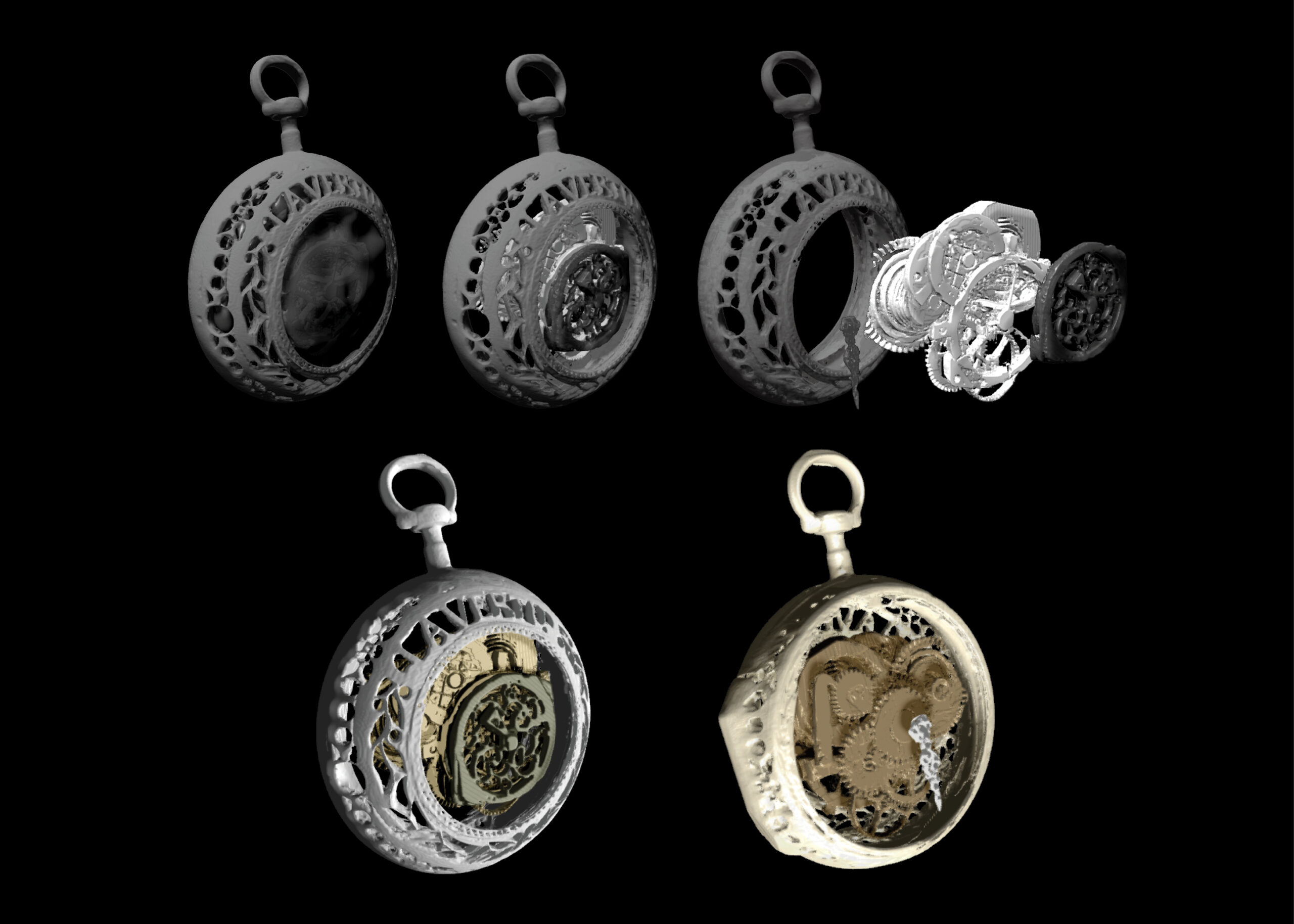
This set of scans of the chatelaine watch has been colorized to show the individual gears and parts using Dragonfly reconstruction software.
Two hours have passed, and the chatelaine watch has now been fully scanned, slice by slice, 1,601 projections in all. Vanderburgh clicks through the images like a flipbook.
The reconstructed 3D rendering is ethereal. Lace-like gears in black, gray and white form concentric circles. (The darker the area, the denser.) From the front, a decorative gear with a filigree pattern is visible. “The watchmakers are artists,” Waddell says, “and have a lot of fun.”
Vanderburgh rotates the image and zooms through from front to back. The two have searched for an engraving.
Alas, there is none.
“There’s some disappointment,” Waddell allows. “It would have been amazing — a stop-the-presses moment for me.” But, she says, that’s no reason to discount Marie Antoinette’s connection to the watch. “There are a lot of links.”
Ditto with the nanoCT. The demo has Waddell’s mind spinning with ideas for exhibitions that combine art and science. On the spot, she imagines displaying the watch with the scans of its insides, perhaps even a 3D-printed version of the mechanism that visitors can pull apart and examine. The collection also has other artifacts that merit a look inside. “There’s lots of opportunities,” she says. “We have tons of miniatures, including a fully functioning piano. It would be really cool to see how it’s put together.”
In the meantime, the demo served its purpose, showcasing the nanoCT’s keen eye.
“You know it’s a watch,” Johnson says. “Suddenly, you can see the gears inside that you couldn’t see before. You understand almost instantly the power of the tool.”
On top of that, the dataset — that 3D reconstruction — can be manipulated. “With your mouse, you can turn the watch around in circles,” he says. “You can flip it over. You can see a cross section. You can pick one gear and get rid of everything else.
“I really love this,” raves Johnson, who could be speaking for any number of nanoCT’s users. “It’s neat.”
Drexel is building its own ‘AWS’ to harness the massive data gleaned from research machines like the nanoCT.
The major scientific instruments operated by Drexel’s Research Core Facilities churn out vast troves of data. Just the X-ray nano-computed tomography (CT) microscope alone produces thousands of high-definition, 3D images per scan.
As of now, all of that raw output is stored locally and must be transported via USB drives, external hard drives, or the cloud to a secondary location for processing on a computer. This is an often slow, cumbersome and expensive undertaking, says Craig Johnson, operations director of Drexel’s Research Core Facilities, the University’s hub of research equipment.
That will change this year. Drexel is building out a new way to automatically and swiftly curate the ever-increasing mounds of research data, including the loads generated by Research Core Facilities’ instruments.
Known as the Platform for Accessible DISE — Data-Intensive Science and Engineering — it will increase storage space eight-fold to 4 petabytes, equal to 4.5 quadrillion bytes or 2 trillion pages of printed text. It will make it easy for scientists in materials science, physics and other areas to transmit their data to an artificial intelligence cluster of high-powered computers, all working together at very high speeds to group similar datapoints and identify patterns. Researchers also will be able to search historical results and train machine-learning models to conduct real-time computations and analyses.
Think of it as Drexel’s own AWS (Amazon Web Services — Amazon’s vast on-demand cloud-computing server network).
“DISE will provide the holy grail to advance information sciences,” says Josh Agar, a former assistant professor at Drexel who wrote a National Science Foundation proposal that resulted in a $4 million award in 2023 to develop special software for scientific workflows.
“We’re really pushing the limits of performance,” he says, “and boundaries of data and research stewardship.” Scientists will be able to ask new research questions, and Drexel will be able to train the next generation of machine learning researchers.
Most clusters, Agar notes, are schedulers, which require waiting in a queue for tasks to get done. DISE will use an orchestration system (the open-source Kubernetes) that allows different applications to run on a set of computers. The resulting automation will lead to better data management, Agar says. The platform also will use a state-of-the-art graphics processing unit to perform calculations — the same way cloud services are done.
DISE, to be based at the University Research Computing Facility hub on Drexel’s University City Campus, is rolling out this year to handle AI workloads. Other more complex aspects may take two to three years to develop. Drexel is collaborating with researchers at Oak Ridge National Laboratory, Morgan State University, and MIT, among others, on the project.
Johnson already sees great potential ahead.
“AI will be really important for analyzing the data that comes out of here,” he says, citing imaging, in situ diagnostics and materials synthesis. “The industry is at the forefront of using AI/machine learning to make new discoveries out of scientific data that weren’t possible before.” — Lini Kadaba